Cardiac Imaging

Cardiac Imaging
- MRI Heart (CMR): creates very detailed images of the heart's anatomy and function, helping in the diagnosis of heart diseases and congenital defects.
- CT Coronary Angiography (CTCA): provides 3D images of the heart and coronary arteries, aiding in the detection of blockages and other issues.
- Coronary Artery Calcium Scoring (CACS): measures the amount of calcium in the walls of the arteries that supply the heart muscle; allows in assessment of the risk of a cardiac event over next 5-10 years.
We are committed to providing comprehensive and affordable Cardiac Imaging to our patients.
FAQs! Need Help?
A coronary artery calcium score is a measurement of the amount of calcium in the walls of the arteries that supply the heart muscle. It is measured by taking a special computed tomography (CT) scan of the heart. The scan shows the amount of hardening of the artery wall (the disease that causes this hardening is called atherosclerosis). The results of the scan make it possible to estimate the risk of a heart attack or stroke (brain attack) in the next 5–10 years. The more calcium (and therefore the more atherosclerosis) there is, the higher the risk of a heart attack or stroke.
A high calcium score does not mean that you will have a heart attack, only that there is a greater likelihood of having one than someone with a low score. Even a person with a score of zero could have a heart attack.
Your doctor will use the coronary artery calcium score to decide whether you are at low, normal or high risk and, if necessary, guide you to reduce your risk. This may be by changes in diet, exercise, controlling blood pressure and diabetes, stopping smoking, and reducing cholesterol in the blood.
This type of scan is a ‘screening’ test; that is, a test you have when you do not have any signs or symptoms of any illness. Screening tests give information about whether a healthy person may have an illness or an increased chance of developing a potentially serious illness. This allows your doctor to provide early advice and, if necessary, treatment before you develop symptoms.
On arrival at the hospital radiology department or private radiology practice, you will be asked to provide your personal details. The radiographer (medical imaging technologist) who will carry out the scan will show you to a changeroom and ask you to put on a gown. You may be asked about your medical history and any medicines you take.
About four electrode patches will be put onto your skin on the front of your chest so an electrocardiogram (ECG) machine can be attached. An ECG machine measures the activity of your heart to show if it is working normally. There will be no injections or drinks given.
You will then be taken to the scanner and asked to lie on a scanning table. The scanner has a round opening and the table moves through the opening during the scan. The ECG machine will be attached to the patches and you can watch the ECG trace of your heart on the monitor. The CT machine links to the ECG so that the recorded electric pulses from your heart tells the CT exactly when to take the scans. You will be asked to hold your breath, the table will move and the pictures of the heart will be taken. The radiographer will check that the scan is a success, and then you can leave.
The scan results will be sent to the doctor who referred you, so you can discuss the score and how it can be used to help you.
There are no after effects. You will be able to carry on your normal day immediately after the scan.
Rarely, skin irritation from the patches used to connect the ECG electrical wires can occur.
The actual CT scan is very quick, but it requires you to hold your breath between 3 and 30 seconds, depending on the individual scanner.
You will need to arrive in time for the radiographer to discuss the scan with you. You will be asked to change into the gown and then you will be set up on the scanner bed. There can be a short delay while the radiographer lets your pulse rate settle if you have been hurrying to the appointment or are nervous. Afterwards, there is a short time while the scan is reviewed to check it is complete, and then you can leave.
You can expect to be in the department for a total of 20–40 minutes.
If you require the result of the scan at the time of your appointment, you can ask the radiology facility when you make the booking. It will take a short time for the CT pictures or images to be processed, reported and made available to you.
The benefit is gaining a better understanding of the relative risk for you of having a heart attack or stroke in the future, and using that information to decide which strategies you should use to reduce your risk if the risk is found to be high.
The calcium score is of no benefit to someone who has already had a heart attack, coronary bypass surgery or a coronary artery stent. These events already indicate a high risk. A calcium score cannot be used to see if any treatment is working or not.
Your doctor may decide that a second calcium score scan after a few years might be helpful to compare the results with the previous scan.
Coronary calcium scores are most informative for women aged between 35 and 70 years and men aged between 40 and 60 years in terms of providing information about cardiovascular risk, or the risk of a heart attack or stroke. Scores in patients outside these age ranges do not have any value in assessing increased risk.
The CT scan is carried out by a radiographer (medical imaging technologist) trained to use the CT scan machine and process the images to measure the amount of calcium in your coronary arteries.
A radiologist (specialist doctor) will look at the result to check the quality of the scan and your medical history, and then write a report for your doctor to use in the management of your heart and atherosclerosis risks.
CTCA is a relatively new test, and the techniques are still evolving with the rapid development of new equipment. There is still disagreement amongst specialist doctors (cardiologists and radiologists) as to the benefits of the test. Published information would suggest that if this test is carried out and no coronary artery disease is detected, your doctor can use this information to manage your symptoms. When the coronary arteries show abnormalities, then your doctor can change your treatment according to the details of the abnormalities shown.
The test has the benefit of being able to show the extent and location of atherosclerosis (a disease that obstructs blood flow in the arteries) within the coronary arteries, even if it is not causing obstruction to the blood flow.
You will be asked to complete a questionnaire before the MRI scan to ensure it is safe for you to enter the MRI machine and be exposed to the magnet.
If you have a history of kidney disease, your doctor might wish to do a blood test before the scan to ensure that the contrast medium (gadolinium) can be safely given, if required (see Gadolinium Contrast Medium for important information for those with impaired kidney function).
No other preparation is required, except for the cardiac stress perfusion MRI where you will be asked to avoid caffeine for 24–48 hours before the test. Caffeine interferes with the action of adenosine (see Stress Perfusion MRI above), which is used to simulate the stress part of this MRI scan. Types of caffeine include tea, coffee, herbal teas, Milo, and even decaffeinated coffee and soft drinks, such as cola. You might also be requested to fast for 6 hours.
Our Services
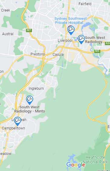
Our Locations
Campbelltown
Building B, Suites 5-9
4 Hyde Parade Campbelltown
Liverpool
Ground Floor, 51 Goulburn St, Liverpool NSW 2170
Minto
North Entrance Car Park
Shop 2A – Minto Mall,
10 Brookfield Road Minto, NSW, 2566
Moorebank
Moorebank Shopping Village
Shop 14C, 42 Stockton Avenue, Moorebank NSW 2170
Wetherill Park
Shop 233 & 235, Stockland Wetherill Park
561 – 583 Polding Street
Wetherill Park NSW 2164
Moorebank Ultrasound Centre
Shop 4, 42 Stockton Ave, Moorebank NSW 2170
Mount Annan
Shop 13-14 Mount Annan Marketplace
11-13 Main Street
Mount Annan NSW 2567
LIVERPOOL
Ground Floor, 51
Goulburn St
Liverpool, NSW, 2170
- M-F: 8.30am - 5.30pm
- Sat: 8:30am - 12:30pm
- Sun: Closed
CAMPBELLTOWN
Park Central, Building B
Ground Level - 4 Hyde Parade
Campbelltown, NSW, 2560
- M-F: 8.30am - 5.30pm
- Sat: 8:30am - 12:30pm
- Sun: Closed
MINTO
North Entrance - Minto Mall
10 Brookfield Road
Minto, NSW, 2566
- M-F: 8.30am - 5.30pm
- Sat: 8:30am - 12:30pm
- Sun: Closed
MOOREBANK
Moorebank Shopping Village
Shop 14C/42 Stockton Avenue
Moorebank, NSW, 2170
- M-F: 8.30am - 5.30pm
- Sat: 8:30am - 12:30pm
- Sun: Closed
MINTO
North Entrance - Minto Mall
10 Brookfield Road
Minto, NSW, 2566
- M-F: 8.30am - 5.30pm
- Sat: 8:30am - 12:30pm
- Sun: Closed
MOOREBANK
Moorebank Shopping Village
Shop 14C/42 Stockton Avenue
Moorebank, NSW, 2170
- M-F: 8.30am - 5.30pm
- Sat: 8:30am - 12:30pm
- Sun: Closed

